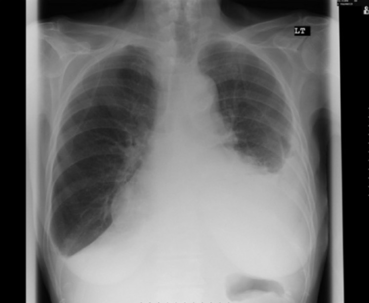
Pleural Effusion Chest X Ray : A left pleural effusion (a) is present ( solid black arrows ).
Pleural Effusion Chest X Ray : A left pleural effusion (a) is present ( solid black arrows ).. A left pleural effusion (a) is present ( solid black arrows ). Pleural effusion is the accumulation of fluid in the pleural space, i.e. Many different types of conditions can cause pleural effusions, with heart failure and pneumonia among the more common ones. The mortality rates associated with pleural e usion seems to be dependent on a variety of factors however, the initial diagnosis of this condition is key for doctors to more e ciently assess the severity and best course 13. Computed tomography (ct) scan of the chest;
This syndrome typically occurs 2 to 3 weeks after a transmural myocardial infarct. A pleural effusion infiltrates the space between these layers. It also can occur following pericardiotomy such as occurs in patients undergoing coronary artery bypass surgery, as in this case. These can show if the. Infection, heart failure, cancer, inflammatory conditions such as lupus, cirrhosis, post heart surgery, pulmonary embolism (clots to the lungs) amongst other causes.
Pleural Effusion Concise Medical Knowledge from cdn.lecturio.com It also can occur following pericardiotomy such as occurs in patients undergoing coronary artery bypass surgery, as in this case. The patient may have unrelated symptoms due to the disease or condition that has caused the effusion.symptoms of pleural effusion include: The lungs and the chest cavity both have a lining that. A left pleural effusion (a) is present ( solid black arrows ). Blunting of the lateral costophrenic angle usually requires about 175 ml but may take as much as 500 ml. A pleural effusion is the accumulation of fluid between the layers of pleura that cover the lung. • g gastric air bubble; These can show if the.
The lungs and the chest cavity both have a lining that.
On physical examination, the patient is stable with decreased breath sounds on the right with dullness to percussion. Pleural effusion is the accumulation of fluid in the pleural space, i.e. About press copyright contact us creators advertise developers terms privacy policy & safety how youtube works test new features press copyright contact us creators. Pleural effusion is a common cause of atelectasis in the adjacent lung. These can show if the. A pleural effusion infiltrates the space between these layers. • p main pulmonary artery; When a person has pleural effusion, it means that fluid has collected in the space between their lungs and chest cavity, or pleural cavity. Plain radiograph chest radiographs are the most commonly used examination to assess for the presence of a pleural effusion; The mortality rates associated with pleural e usion seems to be dependent on a variety of factors however, the initial diagnosis of this condition is key for doctors to more e ciently assess the severity and best course 13. The aetiology of the pleural effusion determines other signs and symptoms. The lungs and the chest cavity both have a lining that. For example, when an infiltrate is accompanied by pleural effusion and other conditions, such as infarction, tuberculosis, infection with mycoplasma, or pneumonia due to hemophilus influenzae, become more likely.
Infection, heart failure, cancer, inflammatory conditions such as lupus, cirrhosis, post heart surgery, pulmonary embolism (clots to the lungs) amongst other causes. Blunting of the lateral costophrenic angle usually requires about 175 ml but may take as much as 500 ml. An increasing number of cases involving pleural fluid that is eventually confirmed to be effusion but not hemothorax. On physical examination, the patient is stable with decreased breath sounds on the right with dullness to percussion. This syndrome typically occurs 2 to 3 weeks after a transmural myocardial infarct.
In Diagnosis Of Pleural Effusion And Pneumothorax In The Intensive Care Unit Patients Can Chest Us Replace Bedside Plain Radiography Sciencedirect from ars.els-cdn.com It also can occur following pericardiotomy such as occurs in patients undergoing coronary artery bypass surgery, as in this case. As little as 5 ml of pleural fluid can be detected by elevating the patient's hips and aiming the central beam at the lateral chest wall parallel to the expected fluid level. Look for consolidation (infection), malignancy, cardiomegaly (cardiac failure) and pleural plaques (asbestos exposure). This syndrome typically occurs 2 to 3 weeks after a transmural myocardial infarct. The lungs and the chest cavity both have a lining that. Infection, heart failure, cancer, inflammatory conditions such as lupus, cirrhosis, post heart surgery, pulmonary embolism (clots to the lungs) amongst other causes. An increasing number of cases involving pleural fluid that is eventually confirmed to be effusion but not hemothorax. Chest films acquired in the lateral decubitus position (with the patient lying on their side) are more sensitive, and can detect as little as 50 ml of fluid.
The diaphragmatic contour is partially or completely obliterated, depending on the amount of the fluid (silhouette sign).
Many different types of conditions can cause pleural effusions, with heart failure and pneumonia among the more common ones. The patient may have unrelated symptoms due to the disease or condition that has caused the effusion.symptoms of pleural effusion include: It also can occur following pericardiotomy such as occurs in patients undergoing coronary artery bypass surgery, as in this case. It may also be referred to as effusion or pulmonary effusion. Pleural effusion is the accumulation of fluid in the pleural space, i.e. Chest films acquired in the lateral decubitus position (with the patient lying on their side) are more sensitive, and can detect as little as 50 ml of fluid. The mortality rates associated with pleural e usion seems to be dependent on a variety of factors however, the initial diagnosis of this condition is key for doctors to more e ciently assess the severity and best course 13. As little as 5 ml of pleural fluid can be detected by elevating the patient's hips and aiming the central beam at the lateral chest wall parallel to the expected fluid level. A pleural effusion is the accumulation of fluid between the layers of pleura that cover the lung. • g gastric air bubble; Normally, the space between the visceral pleura and the parietal pleura cannot be seen. Because the pleural effusion has a density similar to water, it can be seen on radiographs. The aetiology of the pleural effusion determines other signs and symptoms.
Pleural effusion is a common cause of atelectasis in the adjacent lung. Pleural effusions are largely caused by other conditions like cancer. These can show if the. A pleural effusion is the accumulation of fluid between the layers of pleura that cover the lung. A pleural effusion is a buildup of fluid in the pleural space, an area between the layers of tissue that line the lungs and the chest wall.
Pleural Effusion Amboss from media-us.amboss.com • p main pulmonary artery; Pleural effusion is the accumulation of fluid in the pleural space, i.e. For example, when an infiltrate is accompanied by pleural effusion and other conditions, such as infarction, tuberculosis, infection with mycoplasma, or pneumonia due to hemophilus influenzae, become more likely. The diaphragmatic contour is partially or completely obliterated, depending on the amount of the fluid (silhouette sign). Because the pleural effusion has a density similar to water, it can be seen on radiographs. • g gastric air bubble; The fluid may be transude, exudate, blood, chyle or rarely bile. Infection, heart failure, cancer, inflammatory conditions such as lupus, cirrhosis, post heart surgery, pulmonary embolism (clots to the lungs) amongst other causes.
It also can occur following pericardiotomy such as occurs in patients undergoing coronary artery bypass surgery, as in this case.
The fluid may be transude, exudate, blood, chyle or rarely bile. Normally, the space between the visceral pleura and the parietal pleura cannot be seen. Plain radiograph chest radiographs are the most commonly used examination to assess for the presence of a pleural effusion; Blunting of the lateral costophrenic angle usually requires about 175 ml but may take as much as 500 ml. As little as 5 ml of pleural fluid can be detected by elevating the patient's hips and aiming the central beam at the lateral chest wall parallel to the expected fluid level. • p main pulmonary artery; Infection, heart failure, cancer, inflammatory conditions such as lupus, cirrhosis, post heart surgery, pulmonary embolism (clots to the lungs) amongst other causes. On physical examination, the patient is stable with decreased breath sounds on the right with dullness to percussion. For example, when an infiltrate is accompanied by pleural effusion and other conditions, such as infarction, tuberculosis, infection with mycoplasma, or pneumonia due to hemophilus influenzae, become more likely. The aetiology of the pleural effusion determines other signs and symptoms. Look for consolidation (infection), malignancy, cardiomegaly (cardiac failure) and pleural plaques (asbestos exposure). A pleural effusion is the accumulation of fluid between the layers of pleura that cover the lung. Because the pleural effusion has a density similar to water, it can be seen on radiographs.
Related : Pleural Effusion Chest X Ray : A left pleural effusion (a) is present ( solid black arrows )..